This downloadable PDF provides answer keys to cell cycle worksheets, covering topics like interphase, mitosis, and cytokinesis․ It aids in understanding cell division stages and regulation․
Understanding the Cell Cycle
The cell cycle, a fundamental process in all living organisms, governs the growth, replication, and division of cells․ It’s a carefully orchestrated series of events ensuring accurate duplication and distribution of genetic material to create two identical daughter cells․ This cycle is crucial for growth, repair, and reproduction in multicellular organisms, while in unicellular organisms, it serves as the primary means of reproduction․
Understanding the cell cycle involves recognizing its key stages⁚ interphase (G1, S, and G2) and the mitotic phase (mitosis and cytokinesis)․ Interphase is the preparatory phase where the cell grows, replicates its DNA, and prepares for division․ Mitosis, the division of the nucleus, is further divided into prophase, prometaphase, metaphase, anaphase, and telophase, each characterized by specific chromosomal movements․ Cytokinesis, the final step, divides the cytoplasm, resulting in two distinct daughter cells․
Regulation is critical to the cell cycle, ensuring accurate DNA replication and chromosome segregation․ Checkpoints throughout the cycle monitor DNA integrity and proper chromosome duplication, preventing errors that could lead to uncontrolled cell division, such as cancer․ The cell cycle’s intricate mechanisms ensure the faithful transmission of genetic information across generations of cells, maintaining the integrity and continuity of life․
Phases of the Cell Cycle

The cell cycle is a highly ordered sequence of events divided into two main phases⁚ interphase and the mitotic (M) phase․ Interphase, the longer phase, is further subdivided into three distinct stages⁚ G1, S, and G2․ During G1 (Gap 1), the cell grows, synthesizes proteins, and performs its normal functions․ The S (Synthesis) phase is marked by DNA replication, creating an identical copy of each chromosome․ G2 (Gap 2) involves further growth, protein synthesis, and preparation for mitosis․
The M phase encompasses mitosis and cytokinesis․ Mitosis, the division of the nucleus, consists of five stages⁚ prophase, prometaphase, metaphase, anaphase, and telophase․ These stages orchestrate the precise separation of duplicated chromosomes․ Prophase sees chromosome condensation and spindle formation․ Prometaphase involves nuclear envelope breakdown and chromosome attachment to spindle fibers․ Metaphase aligns chromosomes at the cell’s equator․ Anaphase separates sister chromatids to opposite poles․ Telophase involves nuclear envelope reformation and chromosome decondensation․ Finally, cytokinesis divides the cytoplasm, resulting in two separate daughter cells, each with a complete set of chromosomes․ This cyclical process ensures the continuation of life․

Interphase⁚ Growth and Preparation
Interphase, the longest phase of the cell cycle, is a period of intense metabolic activity and growth, preparing the cell for division․ It’s crucial to understand that while seemingly quiescent under a microscope, the cell is actively performing vital functions and undergoing significant internal changes․ This preparatory phase is divided into three distinct stages⁚ G1 (Gap 1), S (Synthesis), and G2 (Gap 2)․ During G1, the cell experiences significant growth, producing proteins and organelles necessary for normal cellular functions․ The cell also accumulates the building blocks required for DNA replication․
The S phase is characterized by DNA replication, where the cell meticulously duplicates its entire genome․ Each chromosome is copied, forming two identical sister chromatids joined at the centromere․ This ensures that each daughter cell receives a complete and accurate set of genetic information․ Following S phase, the cell enters G2, a period of further growth and preparation for mitosis․ The cell synthesizes proteins essential for chromosome manipulation and spindle formation․ Organelles are duplicated, and energy stores are replenished to fuel the energy-demanding process of mitosis․ Interphase culminates in the cell being fully equipped for the complex process of nuclear and cytoplasmic division․
G1 Phase⁚ Cell Growth and Protein Synthesis
The G1 phase, or Gap 1, marks the initial stage of interphase and is a period of substantial cellular growth and metabolic activity․ Fresh from cell division, the cell embarks on a period of intense protein synthesis, producing the enzymes and structural proteins necessary for increasing cell size and carrying out its specific functions․ Organelles are replicated to support the growing cell’s needs, and the cytoplasm expands․ This period is crucial for accumulating the molecular building blocks required for DNA replication in the subsequent S phase, including nucleotides and associated proteins․
Although “Gap” might imply a period of rest, G1 is a dynamic phase filled with biochemical activity․ The cell actively monitors its internal and external environment, ensuring conditions are favorable for proceeding to the next stage of the cell cycle․ Checkpoints operate within G1, assessing DNA integrity and ensuring the cell has sufficient resources to replicate its genome․ If conditions are not met, the cell may enter a non-dividing state called G0, or undergo apoptosis if DNA damage is irreparable․ Successfully navigating G1 sets the stage for the critical process of DNA synthesis in the S phase․
S Phase⁚ DNA Replication
The S phase, or synthesis phase, is the central stage of interphase dedicated to DNA replication․ During this crucial period, the cell meticulously duplicates its entire genome, ensuring each daughter cell receives an identical copy of the genetic material․ This intricate process involves unwinding the double helix, using each strand as a template to synthesize a new complementary strand․ Specialized enzymes, including DNA polymerase, orchestrate this precise duplication, ensuring fidelity and minimizing errors․
As DNA replication progresses, each chromosome transitions from a single chromatid to a pair of identical sister chromatids joined at the centromere․ This duplication doubles the cell’s DNA content without increasing the chromosome number․ The centrosome, responsible for organizing the mitotic spindle, is also duplicated during the S phase, preparing for chromosome segregation in the upcoming M phase․ The successful completion of S phase is critical for maintaining genomic stability and ensuring the accurate transmission of genetic information to the next generation of cells․
G2 Phase⁚ Preparation for Mitosis
The G2 phase, also known as the second gap phase, represents the final stage of interphase, bridging the gap between DNA replication and mitosis․ During G2, the cell undergoes a period of intense preparation, ensuring all necessary components are in place for the complex process of chromosome segregation and cell division․ This preparatory phase involves replenishing energy stores, synthesizing essential proteins required for chromosome manipulation, and duplicating certain cell organelles․
A crucial aspect of G2 is the dismantling of the cytoskeleton, providing the building blocks for the mitotic spindle, the machinery responsible for orchestrating chromosome movement during mitosis․ The cell also undergoes further growth during G2, accumulating the necessary resources to support the energetically demanding process of cell division․ Crucially, G2 includes critical checkpoints that assess the accuracy of DNA replication and ensure any errors are corrected before the cell progresses to mitosis․ This meticulous quality control mechanism helps maintain genomic integrity and prevents the propagation of potentially harmful mutations․
Mitosis (M Phase)⁚ Nuclear Division
Mitosis, a dynamic process of nuclear division, forms the core of the M phase in the cell cycle․ It ensures the precise segregation of duplicated chromosomes into two daughter nuclei, a crucial step in producing genetically identical cells․ This intricate process unfolds through a series of distinct stages⁚ prophase, prometaphase, metaphase, anaphase, and telophase, each marked by specific chromosomal events․
During mitosis, the duplicated chromosomes, initially in a relaxed chromatin state, condense into compact structures, becoming visible under a light microscope․ The nuclear envelope disintegrates, allowing the mitotic spindle, a complex network of microtubules, to interact with the chromosomes․ The spindle fibers attach to specialized protein structures called kinetochores, located at the centromeres of each chromosome․ The chromosomes then align at the cell’s equator, a stage known as metaphase․ Subsequently, the sister chromatids separate and migrate towards opposite poles of the cell, pulled by the shortening spindle fibers․ Finally, the chromosomes decondense, and new nuclear envelopes form around each set, completing the formation of two distinct nuclei․
Prophase⁚ Chromosome Condensation
Prophase, the inaugural stage of mitosis, initiates the dramatic transformation of the cell’s nucleus․ The hallmark of prophase is the condensation of chromatin, the complex of DNA and proteins, into visible chromosomes․ This crucial condensation process organizes and compacts the genetic material, preparing it for the subsequent stages of segregation․ As the chromosomes condense, they become progressively thicker and shorter, taking on their characteristic X-shaped structure, with sister chromatids joined at the centromere․
Simultaneously, within the cytoplasm, the centrosomes, which serve as organizing centers for microtubules, migrate towards opposite poles of the cell․ As they move, they begin to assemble the mitotic spindle, a dynamic network of microtubules that will orchestrate chromosome movement․ The nucleolus, the site of ribosome production, gradually disappears․ Towards the end of prophase, the nuclear envelope starts to break down, dismantling the barrier between the nucleus and cytoplasm․ This breakdown allows the spindle fibers to access and interact with the condensed chromosomes, setting the stage for the next phase of mitosis, prometaphase․ The cell is now fully committed to the process of nuclear division․
Prometaphase⁚ Spindle Attachment
Prometaphase, a dynamic stage of mitosis, bridges the gap between prophase and metaphase․ During prometaphase, the nuclear envelope completely disintegrates, allowing the microtubules of the mitotic spindle to extend into the nuclear region and interact directly with the chromosomes․ These microtubules, emanating from the centrosomes located at opposite poles of the cell, begin to capture and attach to the chromosomes at specialized protein structures called kinetochores, located at the centromeres․
Each chromosome possesses two kinetochores, one on each sister chromatid․ The process of spindle attachment is a dynamic one, with microtubules from both poles vying to connect with the kinetochores․ As microtubules attach and detach, the chromosomes begin to move actively, oscillating back and forth towards the poles․ This “tug-of-war” eventually results in each chromosome becoming bioriented, meaning that its two kinetochores are attached to microtubules emanating from opposite poles․ This biorientation is crucial for the accurate segregation of chromosomes in the subsequent stages of mitosis․ Prometaphase concludes with the chromosomes beginning to align along the metaphase plate, the central plane of the cell․
Metaphase⁚ Chromosome Alignment
Metaphase, a visually striking stage of mitosis, is characterized by the precise alignment of chromosomes along the metaphase plate, the equator of the cell․ This alignment ensures that each daughter cell receives one complete set of chromosomes․ During metaphase, the chromosomes, now maximally condensed and readily visible under a microscope, are held in tension by the opposing forces of the microtubules attached to their kinetochores․
The mitotic spindle, fully formed at this stage, plays a crucial role in maintaining this alignment․ Kinetochore microtubules, connected to the chromosomes, and non-kinetochore microtubules, extending between the poles, work in concert to position the chromosomes precisely․ This delicate balance of forces is essential for the proper segregation of chromosomes in the subsequent stage, anaphase․ Metaphase represents a critical checkpoint in the cell cycle․ The cell verifies that all chromosomes are correctly attached to the spindle and aligned at the metaphase plate before proceeding to anaphase․ This checkpoint ensures the fidelity of chromosome distribution to the daughter cells․ Any errors in attachment or alignment can lead to aneuploidy, an abnormal number of chromosomes, which can have severe consequences for the cell and the organism․
Anaphase⁚ Chromosome Separation
Anaphase, a dynamic stage of mitosis, marks the separation of sister chromatids․ The previously replicated chromosomes, held together at the centromere, are now pulled apart by the shortening kinetochore microtubules․ This separation is triggered by the activation of enzymes that cleave the cohesin proteins holding the sister chromatids together․ As the microtubules retract, each chromatid, now considered an independent chromosome, moves towards opposite poles of the cell․
This movement is facilitated by motor proteins located at the kinetochores, which “walk” along the microtubules, pulling the chromosomes towards the poles․ Simultaneously, the non-kinetochore microtubules elongate, pushing the poles further apart and contributing to the overall separation of the chromosomes․ This precise orchestration ensures that each daughter cell receives a complete and identical set of chromosomes․ Anaphase is a crucial step in ensuring the accurate distribution of genetic material, which is essential for maintaining genomic stability and preventing aneuploidy․ The rapid and coordinated movement of chromosomes during anaphase ensures that the genetic information is equally divided between the two nascent daughter cells․
Telophase⁚ Nuclear Envelope Reformation
Telophase, the final stage of mitosis, essentially reverses the events of prophase and prometaphase․ The chromosomes, having reached their respective poles, begin to decondense, reverting to their less compact chromatin form․ This decondensation allows the genetic material to become accessible again for transcription and other cellular processes․ Simultaneously, the mitotic spindle disassembles, its microtubules breaking down into their tubulin subunits, which can then be reused for other cellular functions, including the formation of the cytoskeleton in the daughter cells․
Crucially, the nuclear envelope begins to reform around each set of chromosomes․ Fragments of the original nuclear membrane, dispersed during prophase, coalesce around the chromatin, eventually forming two distinct nuclear envelopes․ Within these newly formed nuclei, the nucleoli reappear, signifying the resumption of ribosome production․ Telophase marks the completion of nuclear division, setting the stage for cytokinesis, the division of the cytoplasm, which will ultimately produce two separate daughter cells, each with a complete and functional nucleus․ This stage sets the stage for the cell to re-enter interphase and begin a new cycle of growth and division or enter a quiescent state․
Cytokinesis⁚ Cytoplasmic Division
Cytokinesis, the final step in the cell cycle, physically divides the cytoplasm, completing the creation of two independent daughter cells․ This process, distinct from mitosis, typically overlaps with the later stages of mitosis, beginning during anaphase or telophase․ In animal cells, cytokinesis involves the formation of a cleavage furrow, a contractile ring composed primarily of actin filaments․ This ring constricts around the cell’s equator, much like tightening a drawstring, gradually deepening the furrow until the cell membrane pinches inward and separates the two daughter cells․
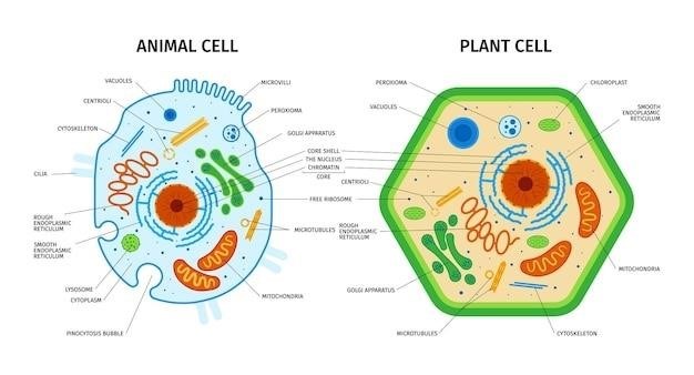
Plant cells, due to their rigid cell walls, undergo cytokinesis differently․ Instead of a cleavage furrow, a cell plate forms between the two newly formed nuclei․ Golgi-derived vesicles containing cell wall materials fuse at the metaphase plate, gradually expanding outward until they reach the existing cell wall, creating a new partition․ This partition develops into a new cell wall, effectively dividing the original cell into two daughter cells, each enclosed by its own membrane and cell wall․ Thus, cytokinesis ensures that each daughter cell receives not only a complete set of chromosomes but also an equal share of cytoplasmic contents, enabling them to function independently․


